


Gonarthrosis of the knee joint is the most common localization of degenerative-dystrophic disease, characterized by gradual destruction of cartilage with subsequent changes in the articular surface, accompanied by pain and decreased mobility.
The disease is more likely to affect women over the age of 40, especially those who are overweight and varicose veins on the lower legs.
The knee joint consists of three compartments:
- medial tibiofemoral;
- tibiofemoral side;
- suprapatellar-femoral.
These compartments can be affected by osteoarthritis deformities (DOA) individually and in any combination. 75% of all cases of gonarthrosis are destruction of the medial tibiofemoral compartment (during movement, it experiences a load exceeding the weight by 2-3 times).
In young patients, only one joint is more often destroyed - right or left (right or left side gonarthrosis).
The cause of DOA knee joint
Several factors may be involved in the development of degenerative cartilage changes simultaneously:
- mechanical load of the knee joint (some specialties, sports) with cartilage microtraumatization;
- as a result of injury, surgical intervention (meniscectomy);
- inflammatory knee disease (arthritis);
- articular surface anatomical inconsistencies (dysplasia);
- static violations (flat feet, curvature of the spine);
- chronic hemarthrosis (accumulation of blood in the synovial cavity);
- metabolic pathology (gout, hemochromatosis, chondrocalcinosis);
- overweight;
- violation of blood supply to the bones;
- osteodystrophy (Paget's disease);
- nerve disease, loss of sensation in the limbs;
- endocrine disorders (acromegaly, diabetes mellitus, amenorrhea, hyperparathyroidism);
- genetic predisposition (a common form of osteoarthritis);
- violation of type II collagen synthesis.
But in 40% of cases, it is impossible to establish the cause of the disease (primary arthrosis).
Pathogenesis of gonarthrosis
early stage
In the early stages of the disease, the metabolic process of cartilage is disrupted. The synthesis and quality of the main structural units of cartilage tissue, proteoglycans, which are responsible for the structural stability of collagen tissues, is reduced.
As a result, chondroitin sulfate, keratin, hyaluronic acid are washed out of the mesh, and the structurally damaged proteoglycans are no longer able to store water. It is absorbed into collagen, a swollen fiber that leads to a decrease in cartilage resistance to stress.
In the synovial cavity, pro-inflammatory substances accumulate, under the influence of which cartilage is destroyed more rapidly. Articular capsule fibrosis develops. Changes in the composition of synovial fluid complicate the delivery of nutrients to the cartilage and affect the sliding of the articular surface during movement.
Pathological progression
In the future, the cartilage gradually becomes thinner, becomes coarse, cracks form throughout its thickness. The bone epiphysis experiences an increased load, which leads to the development of osteosclerosis and the proliferation of compensatory bone tissue (osteophytes).
This body response aims to increase the articular surface area and redistribute the load. But the presence of osteophytes increases discomfort, deformity and in turn limits limb mobility.
Microfractures form in the thickness of the bone, which injure the ducts and lead to intraosseous hypertension. In the last stage of osteoarthritis, the articular surface is completely exposed, deformed, limb movement is very limited.
Symptoms of gonarthrosis of the knee joint
Arthrosis of the knee joint is characterized by a chronic, slowly progressive course (months, years). The clinic developed gradually, with no obvious deterioration. The patient cannot remember exactly when the first symptoms appear.
Clinical manifestations of gonarthrosis:
- sick. Initially, diffuse, short (with long standing, walking up stairs), and as osteoarthritis develops, the pain becomes localized (frontal surface and in the knee), its intensity increases;
- local sensitivity to palpation. Mostly on the inside of the knee along the edge of the joint space;
- Crispy. In stage I it may be inaudible, in stages II-III it accompanies all movements;
- increase in volume, deformity of the knee. As a result of the weakness of the lateral ligaments, a person develops an O -shaped limb configuration (it is clearly visible even in photographs);
- mobility restrictions. At first, there is difficulty with bending the knee, then - with extension.
Causes of pain in DOA:
- mechanical friction of the damaged articular surface;
- increased intraosseous pressure, venous congestion;
- synovitis participation;
- changes in periarticular tissue (stretching of capsules, ligaments, tendons);
- periosteum thickening;
- dystrophic phenomena in adjacent muscles;
- fibromyalgia;
- nerve end compression.
In contrast to coxarthrosis, knee DOA may show spontaneous regression of symptoms.
The clinical manifestations of gonarthrosis depend on the stage:
| Features | I'm on stage | stage II | Level III |
|---|---|---|---|
| sick | Short, occurs more often when the knees are extended (prolonged standing, walking up stairs) | Moderate, disappears after a night's rest | Spoken, annoying even at night |
| Restrictions on mobility | Can not see | There are connection restrictions, mild malfunctions | Continuous bending-extensor contracture, lameness |
| berderai | no | Felt on palpation during movement | crushed away |
| Change the shape | missing | Slight deviation of the limb axis forward, muscle wastage | Valgus or varus deformity. Unstable joints, atrophy of the thigh muscles |
| x-ray images | Slight narrowing of the joint space, early signs of subchondral osteosclerosis | Joint space is narrowed by 50% or more, osteophytes appear | Almost complete absence of joint space, significant deformation and sclerosis of the articular surface, areas of subchondral bone necrosis, osteoporosis |
A frequent complication of knee joint arthrosis is secondary reactive synovitis, which is characterized by the following symptoms:
- increasing pain;
- swollen;
- flow into the synovial cavity;
- increased skin temperature.
Less frequent and more dangerous complications include: joint restriction, osteonecrosis of the femoral condylus, patella subluxation, spontaneous hemarthrosis.
Diagnosis of DOA knee joint
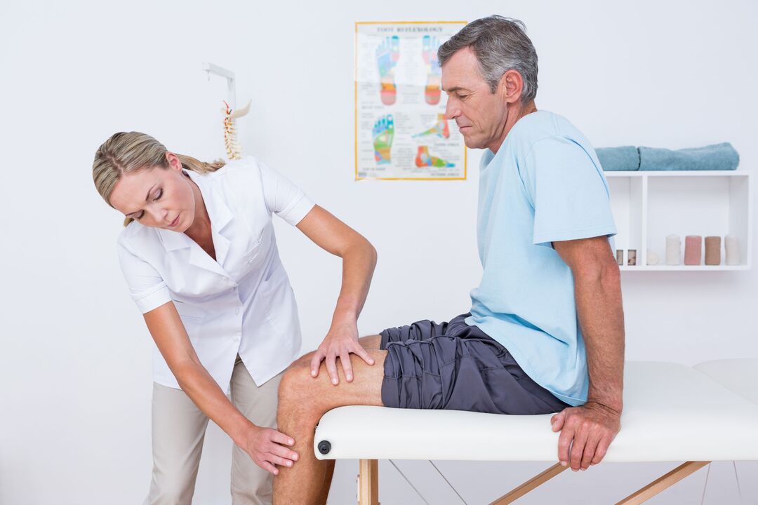
The diagnosis of gonarthrosis is based on the patient’s characteristic complaints, changes detected during the examination and the results of additional tests.
To confirm osteoarthritis, it is prescribed:
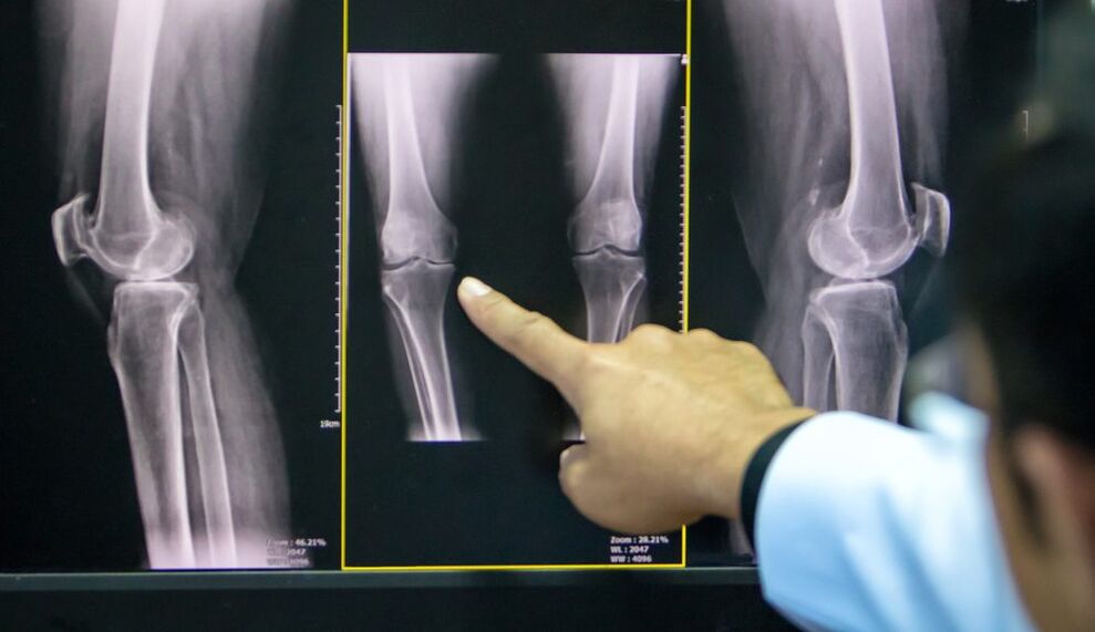
- radiography of the knee joint in two projections (anteroposterior and lateral): the easiest way to confirm the diagnosis at an advanced stage of pathology;
- Ultrasound: determination of the presence of effusions in the joints, measurement of cartilage thickness;
- synovial fluid analysis;
- diagnostic arthroscopy (visual assessment of cartilage) with biopsy;
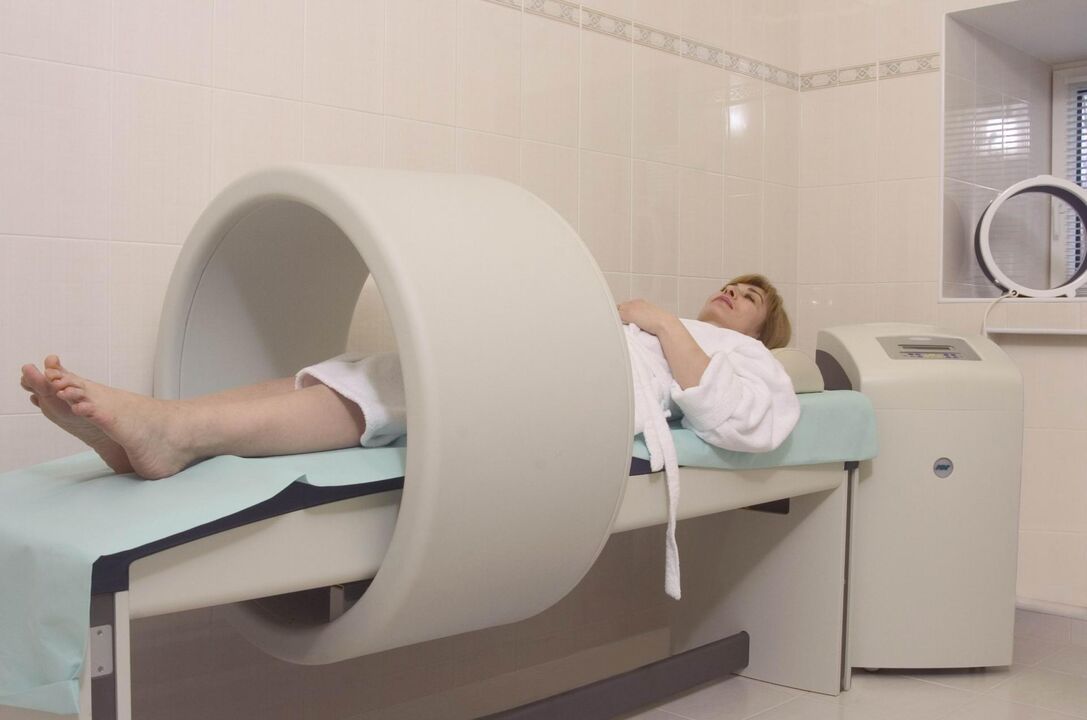
- computation and magnetic resonance imaging (CT, MRI): the best method for diagnosing DOA at an early stage.
If the doctor has doubts about the diagnosis, it may be prescribed:
- scintigraphy: scanning the joints after the introduction of radioactive isotopes;
- thermography: the study of the intensity of infrared radiation (its intensity is directly proportional to the intensity of inflammation).
Treatment of gonarthrosis of the knee joint
The treatment regimen for osteoarthritis combines several approaches: non -drug methods, pharmacotherapy and surgical correction. The ratio of each method was determined individually for each patient.
Non -drug treatment
In the latest ESCEO (European Association for Clinical Aspects of Osteoporosis and Osteoarthritis) guidelines on how to treat knee osteoarthritis, experts place special emphasis on patient education and lifestyle modifications.
Patients need:
- explain what the essence of the disease is, prepared for long -term treatment;
- teach how to use aids (sticks, orthoses);
- prescribing a diet (for patients with a body mass index over 30);
- provide a set of exercises to strengthen the thigh muscles and unload the knee joint;
- explains the importance of increasing physical activity.
In the early stages of knee arthrosis, physiotherapy treatment methods give good results:
- massage;
- magnetotherapy;
- UHF therapy;
- electrophoresis;
- hydrogen sulfide bath;
- paraffin application;
- acupuncture.
Pharmacotherapy of gonarthrosis
The use of drugs in DOA is intended to relieve pain, reduce inflammation, and slow the rate of cartilage destruction.
Symptomatic treatment:
- analgesic;
- non-steroidal anti-inflammatory substances (NSAIDs) of the COX-2 inhibitor group in the form of tablets or suppositories;
- non -narcotic analgesics (with resistant pain syndrome).
Structural modifiers (chondroprotectors):
- Chondroitin sulfate;
- Glucosamine sulfate.
These drugs can be taken in capsule form in a course several times a year, injected intramuscularly or directly into the synovial cavity.
Local therapy includes close and intra-articular injections of glucocorticosteroids, hyaluronic acid preparations.
At stages I – II of DOA, an important place in complex therapy is the use of anti-inflammatory ointments, gels and creams based on NSAIDs. They help reduce a patient’s need to take NSAIDs orally, thus reducing the risk of damage to the digestive tract.
Restoration of the people
The use of tinctures, decoctions, extracts, local application of medicinal plants should be considered as an additional method for the treatment of DOA, folk remedies should not replace the therapy prescribed by a doctor.
Plants used in osteoarthritis: dandelion, ginger, Jerusalem artichoke, burdock, garlic, sea buckthorn.
Surgery
Surgical intervention may be required at all stages of gonarthrosis with the effect of inadequate medical measures. The most common is an endoscopic procedure, in the most severe cases, endoprosthesis replacement is indicated.
Type of endoscopic intervention:
- revision and restoration of joints: extraction of inflammatory contents from the synovial cavity, cartilage fragments;
- plasma or laser ablation: removal of mechanical obstructions in the synovial cavity;
- chondroplasty.
Corrective periarticular osteotomy is indicated for patients with early manifestations of axial limb deformity (no more than 15-20%).
The purpose of the operation is to restore the normal configuration of the joint, distribute the load evenly over the articular surface, and remove the damaged area. This procedure allows you to delay arthroplasty.
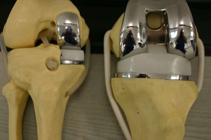
Instructions for replacing the affected area (or entire joint) with an artificial one:
- DOA Degree II-III;
- severe axial deformity of the limb;
- aseptic necrosis of the subchondral bone layer;
- persistent pain syndrome.
Contraindications to knee arthroplasty:
- total damage to the joints;
- unstable ligament apparatus;
- DOA due to inflammatory arthritis;
- persistent flexural contractions, severe muscle weakness.
In this case, the patient underwent arthrodesis - a comparison of the knee joint in physiological position with removal of the articular surface. This relieves pain but shortens the leg, causing secondary lesions on the knee, hip and contralateral spine.
Prevention
Prevention of premature cartilage degeneration should begin in childhood.
Precautions:
- prevention of scoliosis;
- flat foot correction (shoes with arch support);
- regular physical education (limit heavy sports);
- exclusion of fixed posture while working.